Page 171 - Kỷ yếu hội thảo quốc tế: Ứng dụng công nghệ mới trong công trình xanh - lần thứ 9 (ATiGB 2024)
P. 171
162                              TRƯỜNG ĐẠI HỌC SƯ PHẠM KỸ THUẬT - ĐẠI HỌC ĐÀ NẴNG

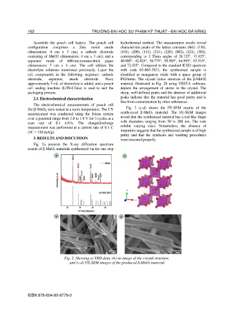
                  Assemble  the  pouch  cell  battery.  The  pouch  cell   hydrothermal  method.  The  measurement  results  reveal
               configuration  comprises  a  Zinc  metal  anode   characteristic peaks of the lattice constants (hkl): (110),
               (dimensions:  4  cm  x  3  cm),  a  cathode  electrode   (101),  (200),  (111),  (211),  (220),  (002),  (221),  (301),
               consisting of MnO2 (dimensions: 4 cm x 3 cm), and a   corresponding  to  2  Theta  angles  of  28.725°,  37.425°,
               separator  made  of  600-micrometer-thick  paper   40.905°,  42.825°,  56.775°,  59.505°,  64.995°,  67.515°,
               (dimensions:  5  cm  x  4  cm).  The  cell  utilizes  the   and 72.435°. Compared to the standard ICSD spectrum
               electrolyte  solutions  mentioned  previously.  Layer  the   with  code  03-065-2821,  the  synthesized  sample  is
               cell  components  in  the  following  sequence:  cathode   identified  as  manganese  oxide  with  a  space  group  of
               electrode,   separator,   anode   electrode.   Next,   P42/mnm.  The  crystal  lattice  structure  of  the  β-MnO2
               approximately 5 mL of electrolyte is added, and a pouch   material, illustrated in Fig. 2b using VESTA  software,
               cell  sealing  machine  (LiTh-China)  is  used  to  seal  the   depicts  the  arrangement  of  atoms  in  the  crystal.  The
               packaging process.                             sharp, well-defined peaks and the absence of additional
                  2.3. Electrochemical characterization       peaks indicate that the material has good purity and is
                                                              free from contamination by other substances.
                  The  electrochemical  measurements  of  pouch  cell
               Zn//β-MnO 2 were tested at a room temperature. The CV   Fig.  2  (c-d)  shows  the  FE-SEM  results  of  the
               measurement  was  conducted  using  the  Ivium  system   synthesized  β-MnO2  material.  The  FE-SEM  images
               over a potential range from 1.0 to 1.8 V for 3 cycles at a   reveal that the synthesized material has a rod-like shape
               scan  rate  of  0.1  mV/s.  The  charge/discharge   with  diameters  ranging  from  50  to  200  nm.  The  rods
               measurement was performed at a current rate of 0.1 C   exhibit  varying  sizes.  Nonetheless,  the  absence  of
               (1C = 310 mA/g).                               impurities suggests that the synthesized sample is of high
                                                              purity  and  that  the  synthesis  and  washing  procedures
                  3. RESULTS AND DISCUSSION                   were executed properly.
                  Fig.  2a  presents  the  X-ray  diffraction  spectrum
               results of β-MnO 2 materials synthesized via the one-step


                                  Fig. 2. Showing a) XRD data, (b) an image of the crystal structure,
                                    and (c-d) FE-SEM images of the produced β-MnO2 material


               ISBN:978-604-80-9779-0

